
Please use this identifier to cite or link to this item:
http://repo.knmu.edu.ua/handle/123456789/3556Full metadata record
| DC Field | Value | Language |
|---|---|---|
| dc.contributor.author | Honchar, Oleksii | - |
| dc.contributor.author | Гончарь, Олексій Володимирович | - |
| dc.contributor.author | Гончарь, Алексей Владимирович | - |
| dc.contributor.author | Kovalyova, Olga | - |
| dc.contributor.author | Ковальова, Ольга Миколаївна | - |
| dc.contributor.author | Ковалёва, Ольга Николаевна | - |
| dc.contributor.author | Demydenko, Ganna | - |
| dc.contributor.author | Демиденко, Ганна Валеріївна | - |
| dc.contributor.author | Демиденко, Анна Валерьевна | - |
| dc.date.accessioned | 2013-06-26T05:36:29Z | - |
| dc.date.available | 2013-06-26T05:36:29Z | - |
| dc.date.issued | 2013 | - |
| dc.identifier.citation | Honchar O. Diastolic dysfunction, left ventricular and vascular remodeling in hypertensive patients with obesity / О. Honchar, O. Kovalyova, G. Demydenko // Journal of Hypertension. – 2013. – Vol. 30, e Supplement A : 23rd European Meeting on Hypertension and Cardiovascular Protection, Milan, 14-17 June 2013 : Abstracts. – Р. e347. | uk_UA |
| dc.identifier.uri | https://repo.knmu.edu.ua/handle/123456789/3556 | - |
| dc.description.abstract | Objective. To investigate the peculiarities of left ventricular (LV) remodeling, diastolic dysfunction (DD) and common carotid artery (CCA) remodeling in hypertensive patients with obesity. Design and method. 75 hypertensive patients (32 male, 43 female) with preserved LV systolic function had been observed, including 51 obese patients (21 male, 30 female). An ultrasound examination of heart (including estimation of transmitral blood flow and mitral valve annulus motion parameters) and CCA was performed. LV geometric pattern, E/A and E/Em ratios, ring segment weight (VM) of CCA were calculated. The statistical analysis was conducted using Mann-Whitney and Pearson χ2 methods. Results. Normal LV geometrical pattern was observed in 6 (11,8%) obese patients and 4 (16,7%) – without obesity, p>0,05; concentric remodeling – 7 (13,7%) and 1 (4,2%), p>0,05; concentric hypertrophy (CH) – 29 (56,9%) and 8 (33,3%), р=0,028; eccentric hypertrophy – (EH) 9 (17,6%) and 10 (41,7%), р=0,015. LV CH prevalence in obese patients was significantly higher compared to EH, p<0,0001. Patients with obesity had higher LVMM (309,3±15,9 vs 258,8±16,8 g, p<0,05), but not MMI (148,5±7,1 vs 139,7±7,9 g/m2, p>0,05). LV DD was revealed in 48 (94,1%) obese patients (including 23 (100%) with II-III st. obesity) and 19 (79,1%) – without obesity, p=0,05. Type I of DD was observed in 31 (60,8%) obese patients, type II – in 17 (33,3%), p=0,028; in non-obese patients – 12 (50,0%) and 7 (29,2%) accordingly, p=0,07. CCA wall hypertrophy was observed in 44 (86,3%) obese patients (including 22 (95,7%) with II-III st. obesity) and 18 (75,0%) – without obesity, p<0,01. The values of mitral valve annulus motion parameters, intimal medial thickness and ring segment weight of CCA are given below as median [LQ; UQ]. Conclusion. Hypertensive patients with obesity were characterized by higher prevalence of LV hypertrophy, concentric patterns of remodeling and DD. Indices of CCA remodeling were also increasing along with BMI. Tissue Doppler parameters of mitral valve annulus motion are important early markers of LV DD. | uk_UA |
| dc.language.iso | en | uk_UA |
| dc.subject | essential hypertension | uk_UA |
| dc.subject | hypertensive | uk_UA |
| dc.subject | obesity | uk_UA |
| dc.subject | obese | uk_UA |
| dc.subject | myocardial remodeling | uk_UA |
| dc.subject | left ventricular remodeling | uk_UA |
| dc.subject | diastolic dysfunction | uk_UA |
| dc.subject | vascular remodeling | uk_UA |
| dc.subject | arterial stiffness | uk_UA |
| dc.subject | elastic properties | uk_UA |
| dc.subject | common carotid arteries | uk_UA |
| dc.subject | гіпертонічна хвороба | uk_UA |
| dc.subject | артеріальна гіпертензія | uk_UA |
| dc.subject | ожиріння | uk_UA |
| dc.subject | ремоделювання міокарда | uk_UA |
| dc.subject | ремоделювання лівого шлуночка | uk_UA |
| dc.subject | діастолічна дисфункція | uk_UA |
| dc.subject | ремоделювання судин | uk_UA |
| dc.subject | жорсткість | uk_UA |
| dc.subject | пружно-еластичні властивості | uk_UA |
| dc.subject | загальні сонні артерії | uk_UA |
| dc.subject | гипертоническая болезнь | uk_UA |
| dc.subject | артериальная гипертензия | uk_UA |
| dc.subject | ожирение | uk_UA |
| dc.subject | ремоделирование миокарда | uk_UA |
| dc.subject | ремоделирование левого желудочка | uk_UA |
| dc.subject | диастолическая дисфункция | uk_UA |
| dc.subject | ремоделирование сосудов | uk_UA |
| dc.subject | жёсткость | uk_UA |
| dc.subject | упруго-эластические свойства | uk_UA |
| dc.subject | общие сонные артерии | uk_UA |
| dc.title | Diastolic dysfunction, left ventricular and vascular remodeling in hypertensive patients with obesity | uk_UA |
| dc.title.alternative | Діастолічна дисфункція, ремоделювання лівого шлуночка та судин у хворих на гіпертонічну хворобу з ожирінням | uk_UA |
| dc.title.alternative | Диастолическая дисфункция, ремоделирование левого желудочка и сосудов у больных гипертонической болезнью с ожирением | uk_UA |
| dc.type | Thesis | uk_UA |
| Appears in Collections: | Наукові праці. Кафедра пропедевтики внутрішньої медицини № 1, основ біоетики та біобезпеки | |
Files in This Item:
| File | Description | Size | Format | |
|---|---|---|---|---|
| Гончарь О.В._2013_007.pdf | Тези конференції | 230,46 kB | Adobe PDF |  View/Open |
| Гончарь О.В._2013_007.doc | Препринт | 41 kB | Microsoft Word | View/Open |
| 1.jpg | Таблиця | 187,53 kB | JPEG | 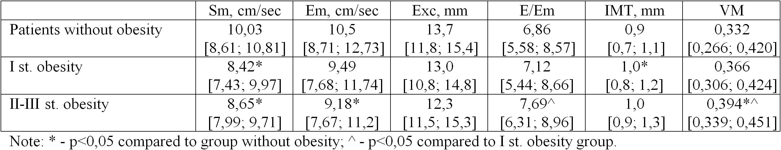 View/Open |
Items in DSpace are protected by copyright, with all rights reserved, unless otherwise indicated.